1000 AI Algorithms in Medical Imaging – Thoughts for Tech Entrepreneurs (1 of 5)
In the spirit of graduation season, I’d like to share some thoughts with the new wave of tech entrepreneurs interested in AI. In this weekly 5 part series, I will lay out a little background in the industry and explain current workflow and pain points. There is a great benefit coming into this field of AI in medical imaging now as we have learned a ton in the past decade and are ready for the next wave of enthusiastic, innovative, hard working and caring leaders to build great platforms for patients and providers. If/when you create an awesome platform, it may just help you or your family down the road.
From high school graduation to completing a radiology fellowship takes about 14 years. With nano degrees and other resources, a creative entrepreneur could spend a third of that time, or less, building great products for the health system if you leverage available resources properly. These resources include current people in the very large health system and I will get into that in article 2.
You don’t need specific medical knowledge for this series, I will explain processes and medical concepts at broader levels and if any of them sound like good problems to solve, you will be armed with enough information to begin your search to dig in more. Importantly, in article 3, I will explain that just finding things on an image from a publicly available dataset may not be a reasonable plan anymore and this is explained nicely by Dr. Harvey in this blog post.
Also in article 3, I will discuss that deploying a health solution is not like adding an extension to your Chrome browser. At this time, it is more than that. By knowing a few places to land, you can build a better plan.
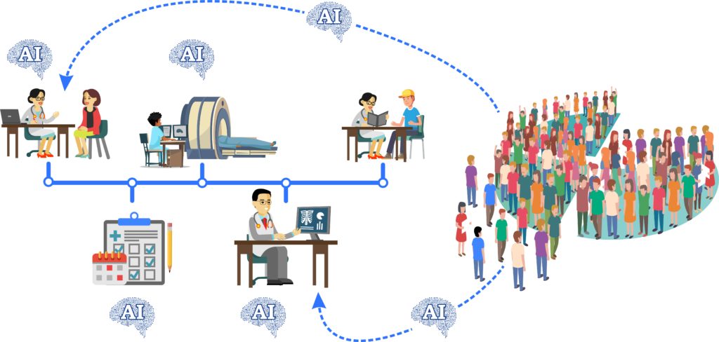
In article 4, I’ll give some ideas on how radiology fits in a larger clinical pathway affecting specific patients. Here is an example: A patient with heart failure uses medicine to help them rid fluid in urine to prevent extra fluid in the lungs. Using a bathroom scale can estimate how much fluid is being accumulated in this patient’s body. How can we connect data from a smart bluetooth scale in the home of a patient with congestive heart failure with data from a radiograph if they present to the emergency department with shortness of breath? What are the variables to consider to determine if this is a good problem to solve?
Article 5 will discuss systemic issues. There are specific problems across the entire health system that deserves a new process. Two examples are: fax machines and clipboards. I will end with an introduction of the most important piece of information if you intend on selling your product, regulatory processes to include FDA evaluation.
Some of you will be scared off by these articles and that may be for the best. If this was easy, everyone would do it and there would be no problems to solve. So to the few, the tough, the innovators let’s go.
Within the past 10 years, nearly 70 AI algorithms have been approved by the FDA for medical imaging. This has been an impressive display of innovation and creativity to get to this point however I’d like to place this in context.
Dr. Charles Khan is a long time leader in radiology and if we use his published numbers, there are over 12,000 medical conditions that cause findings. This means for most radiologists, the current AI platforms cover an insignificant part of their work. For some it is gaining traction, particularly in large teleradiology groups where smart work lists and triage algorithms are making a real improvement. For fewer radiologists still, there are the academic pioneers who use this every day and see mind boggling results in not only imaging, but clinical data as well.
Dr. Khan has painstakingly curated and noted over 4000 imaging findings. Just to clarify a ‘finding’ can be many things. The presence or absence of something, or increase or decrease in size, or change in appearance can be a finding. Many of these imaging findings are perfectly suitable for training with a properly sized data set. Another point to consider – these are findings that we can physically see with human eyes.
Data is collected from CT and MRI scanners and manipulated quite a bit so human eyes and brains can make sense of it. With that, AI should be able to capture data in images, and from source data, that was never clinically evaluated previously. So this number 4000 has nowhere to go but up. Also remember, there are things algorithms can do much faster such as precise measuring irregular brain lesions or pulmonary nodules.
With 12,000 medical conditions and 4000 imaging findings in the field of radiology, the first 70 algorithms are a great start but there is room to innovate. I picked 1000 as a goal. Let’s see how long it takes for the next 930 FDA approved AI algorithms. Will one of them be yours?
In the next article I will explain the steps and subsequent data collected during the clinical process. I will start from radiology exam ordering and scheduling to viewing and interpreting and discuss why it is important to have this background to make the most useful AI application.